
Researchers identified two brain areas in mice that helped the animals learn to suppress their instinctive fears of predators
Sara Hashemi February 11, 2025
Humans have certain instinctive fear reactions to things that won’t actually harm us—we might jump at a too-loud noise or shriek at a spider, for example. But what if we could overwrite our instincts?
Neuroscientists have figured out which regions of the brain suppress these fear responses in mice, a finding they say could one day help people with phobias, anxiety and post-traumatic stress disorder. Their results were published in the journal Science last week.
“We’ve uncovered the mechanism by which the brain—through experience—can understand which potential instinctive dangers are actually not a danger,” says lead author Sara Mederos, a neuroscientist at University College London’s Sainsbury Wellcome Center, to the Washington Post’s Leo Sands.
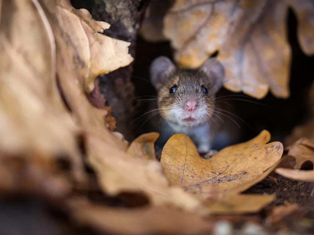
Mederos and her team repeatedly exposed around 100 mice to one of their instinctive fears: the shadow of a bird. At first, the mice would hide at the perceived threat. Then, the researchers installed a barrier preventing the mice’s escape. With repeated exposure, the mice eventually learned that the shadow posed no danger to them, and they remained calm. Even once the team removed the barrier, the mice largely did not attempt to flee.
“I like their behavioral model,” says Christina Perry, a behavioral neuroscientist at Macquarie University in Australia who was not involved in the study, to Felicity Nelson at Nature News. “It’s very simple,” she adds. The mice “don’t get eaten, so they learn that this fake predator is not, in fact, a threat.”
In some cases, while the mice were exposed to the shadow, the researchers silenced specific neurons in their brains through a technique called optogenetics. The study found two parts of the brain that played a role in suppressing visual fears. One was the visual cortex, which appeared to be key in learning that certain things were not a threat. The other part was unexpected—called the ventrolateral geniculate nucleus (vLGN), it was found to store memories related to ignoring fears.
/https://tf-cmsv2-smithsonianmag-media.s3.amazonaws.com/filer_public/bf/e2/bfe2be12-2508-4c65-9dbe-3f1c76d101ce/coronal_brain_slice_final_website.png)
“Our results challenge traditional views about learning and memory,” says Sonja Hofer, a neuroscientist at the Sainsbury Wellcome Center who led the research with Mederos, in a statement. The cerebral cortex, where the visual cortex is located, has long been considered the brain’s center for “learning, memory and behavioral flexibility,” she adds.
“We did not know that there is a chance of plasticity and learning happening in these downstream areas,” says Mederos to the Washington Post. But when the vLGN was switched off in mice that had already learned the shadow was non-threatening, those same animals reacted to the shadow with fear.
“It is really one of the few examples where changes in the brain have been directly linked to changes in behavior following experience,” says Alexander Heimel, a scientist at the Netherlands Institute for Neuroscience who was not involved in the study, to Sydney Wyatt at the Transmitter. “It was a big surprise to me that the site of the plasticity turned out to be the vLGN.”
The scientists believe that their findings could advance treatment around fear-related disorders.
“Our findings could also help advance our understanding of what is going wrong in the brain when fear response regulation is impaired in conditions such as phobias, anxiety and PTSD,” Hofer says in the statement. “While instinctive fear reactions to predators may be less relevant for modern humans, the brain pathway we discovered exists in humans too.” Still, the team notes that any development like this wouldn’t take place until the far future.